As previously discussed, tumors are named for the tissue they originate from. Gliomas are tumors made from glial cells. Most likely, a neurosurgeon will obtain brain tissue during surgery to make an accurate diagnosis and to guide treatment options. Size and location of a tumor are important factors when learning how the brain may be affected by a glioma. Please check out the pictures below for a better understanding.
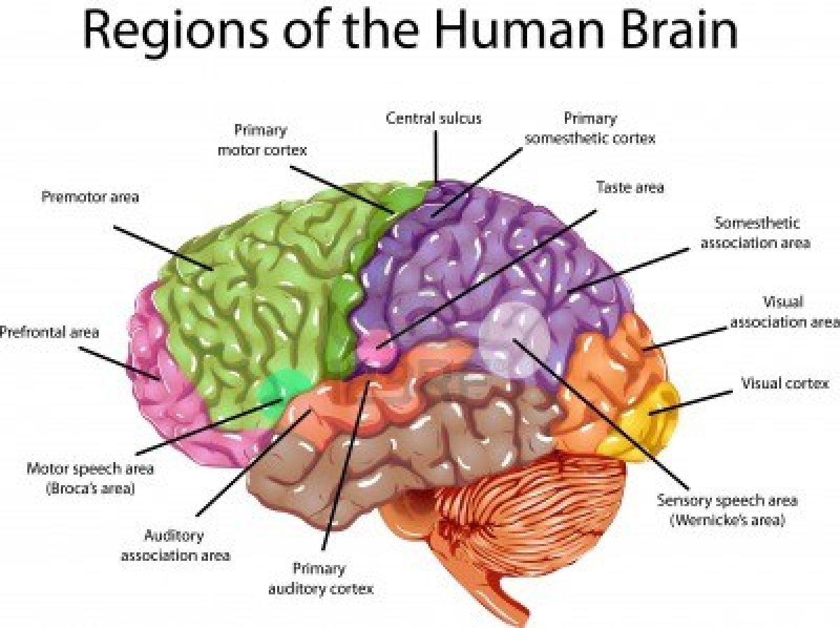 Each area of the brain is color-coded. If a tumor were to form, the corresponding functions would be affected. |
 This model shows how sensory and motor (movement) would be affected. Keep this in mind: if a tumor forms on the left side of the brain, the right side of the body will experience sensory and/or motor deficits. |
| Source: https://classconnection.s3.amazonaws.com/917/flashcards/770260/png/homunculus.png
In the case of gliomas, the progression of disease is quite dismal. Most gliomas are considered to be malignant and therefore patients do not usually have a favorable outcome. Again, it is dependent upon the location, size and type of brain tumor. Recall that gliomas can be any of these types: astrocytoma, glioblastoma multiforme, oligodendroglioma, ependymoma or medulloblastoma; all of which are malignant except the oligodendroglioma (benign tumor). Medical-Surgical Nursing reports a 5-year survival rate after the diagnosis of a primary brain tumor (1375).
Sources:
Homunculus: https://classconnection.s3.amazonaws.com/917/flashcards/770260/png/homunculus.png
Lewis, S., Dirksen, S., Heitkemper, M., & Bucher, L. (2014). Nervous System. In Medical-Surgical Nursing: Assessment and Management of Clinical Problems (9th ed., p. 1377). St. Louis: Elsevier Mosby.
|
No comments:
Post a Comment